What Is a Transthoracic Echocardiogram? A Simple Guide for Patients
What is an Echocardiogram?
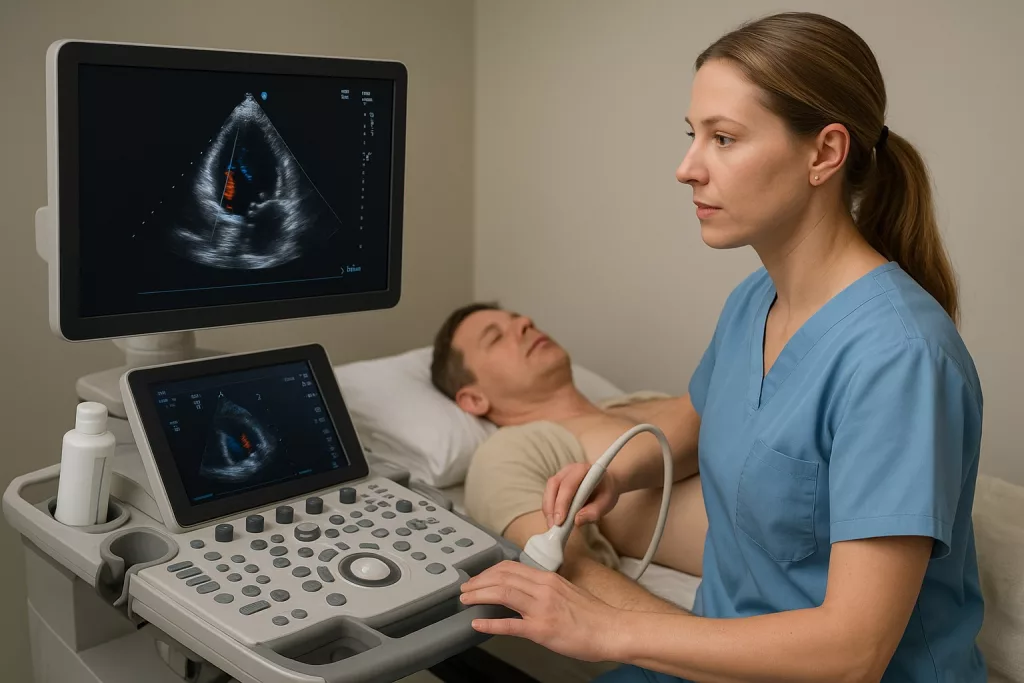
An echocardiogram is a non-invasive ultrasound examination that uses high-frequency sound waves to produce detailed, real-time images of the heart’s structures and function. It allows us to visualise the heart chambers and valves to assess how effectively the heart is pumping blood throughout the body.

Why Might You Need an Echocardiogram?
Your doctor may refer you for an echocardiogram to:
- Check the overall health of your heart.
- Assess heart muscle function, for example after a heart attack or in suspected heart failure
- Detect and monitor structural heart diseases, such as valve stenosis or regurgitation.
- Investigate symptoms like chest pain, shortness of breath, or palpitations.
- Investigate congenital (present from birth) heart abnormalities.
- Monitor an existing heart condition.
- Check for fluid around the heart (pericardial effusion) or blood clots within the heart chambers.
Preparing for Your Echocardiogram
- No special preparation is needed for a standard echocardiogram. You can eat and drink normally.
- Please wear comfortable, loose-fitting clothing. You may be asked to remove clothing from the waist up and wear a gown.
- Bring a list of any medications you are currently taking.
- If you have any mobility restrictions or special requirements, please inform us prior to your appointment so we can make suitable arrangements.
What to Expect During the Procedure
- The scan is typically performed in a dedicated cardiac ultrasound room and takes approximately 30 minutes.
- You will be asked to lie on an examination couch, usually positioned on your left side to bring the heart closer to the chest wall for optimal imaging.
- A water-based ultrasound gel will be applied to your chest to ensure good contact between the skin and the transducer (ultrasound probe).
- A small amount of cool gel will be applied to your chest — this helps the ultrasound probe make good contact with your skin.
- You may hear pulsed or whooshing sounds — these are Doppler signals, which allow us to assess blood flow through the heart and valves.
- You may be asked to briefly hold your breath or change position to improve image quality.
- The procedure is painless, although you may feel mild pressure as the probe is moved across your chest.
After the Examination
- There is no recovery time needed — you can go straight back to your normal activities, including driving.
- The images will be reviewed after the scan and the report will be sent to the doctor who referred you.
- Your referring cardiologist will discuss the results with you at your next appointment.
Are There Any Risks?
A standard echocardiogram is very safe. It uses sound waves, not radiation. There are no known risks or side effects. Rarely, minimal discomfort may be experienced due to the probe pressure, but this is temporary.
Author’s biography

Ashly Johnson
Senior Cardiac physiologist
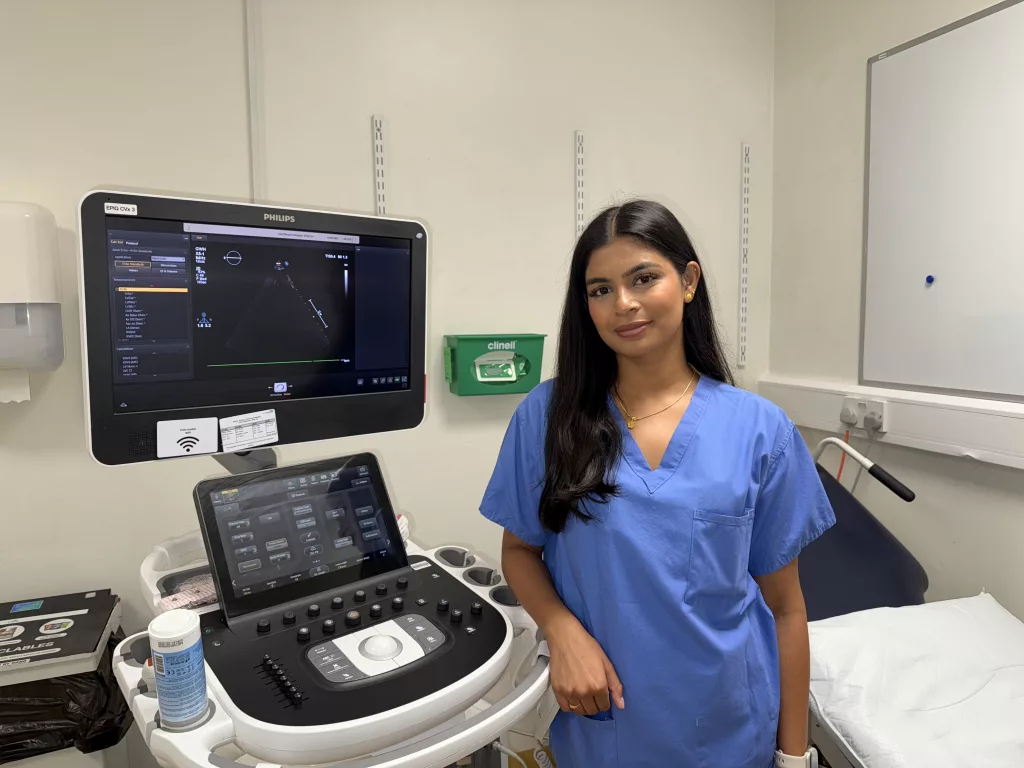
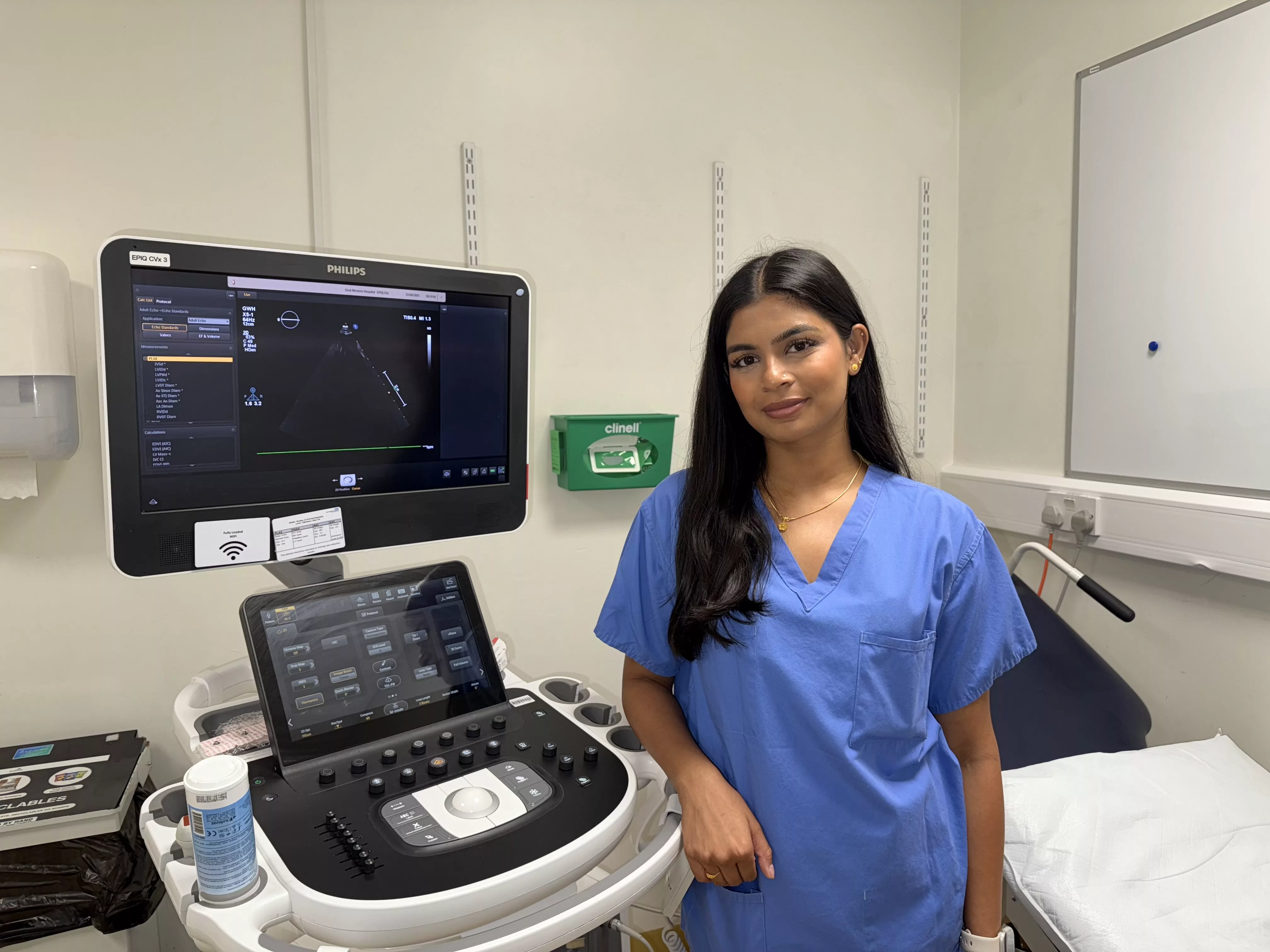
I am a highly specialist cardiac physiologist with British Society of Echocardiography (BSE) accreditation, achieved through the Echo Training Program. Since qualifying in 2021, I have developed extensive experience in performing and interpreting transthoracic echocardiograms, supporting the diagnosis and management of a wide range of cardiovascular conditions. I am committed to maintaining high clinical standards and continuing professional development to ensure the best possible care for patients.


Leave feedback about this